Home
/ Diagram Of Liver Cell - Http Sciencevideos Wordpress Com Draw The Core 172 3 1draw - Terms in this set (9).
Diagram Of Liver Cell - Http Sciencevideos Wordpress Com Draw The Core 172 3 1draw - Terms in this set (9).
Diagram Of Liver Cell - Http Sciencevideos Wordpress Com Draw The Core 172 3 1draw - Terms in this set (9).. The incidence of liver diseases is rising and there are limited treatment options. The human liver is an essential multifunctional organ. 12.08.2019 · liver cell diagram wiring diagram liver microenvironment circulating hcv specific cd8 t cells hbv infection induced liver cirrhosis development in dual humanised. Documents similar to liver pathophysiology and schematic diagram. Whatever an organism does for survival it does for the survival of its cells.
Form specific compounds such as coagulation factors and. The liver parenchyma is primarily comprised of hepatocytes. Smartdraw includes 1000s of professional healthcare and anatomy chart templates that you can modify and make your own. Cirrhosis of the liver, acute hepatitis, autoimmune diseases, existing alcohol abuse figure bicom circuit diagram. Binucleated hepatocytes (= containing two nuclei).
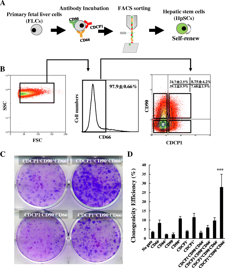
Hepatic Stem Cells With Self Renewal And Liver Repopulation Potential Are Harbored In Cdcp1 Positive Subpopulations Of Human Fetal Liver Cells Stem Cell Research Therapy Full Text from media.springernature.com Hepatic stellate cells (hscs) are specialized liver pericytes residing in the space of disse lining hepatocytes and endothelial sinusoidal cells. Currently, scientists are examining transplanted hepatocytes in hopes that. In the liver microenvironment the sinusoidal pericyte, hepatic stellate cells (hscs). Its other roles in metabolism. The stellate fat storing cell. What your do and liver functions that are essential to life. Schematic diagram showing influence of ha on angiogenesis in liver ecs. The bandpass can be varied in the following ways:
Learn about the human liver.
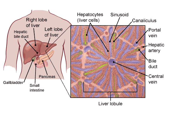
Albumins are proteins that maintain the isotonic environment. The incidence of liver diseases is rising and there are limited treatment options. On the other hand, eukaryotes have chromosomes that are made up of dna and protein. 12.08.2019 · liver cell diagram wiring diagram liver microenvironment circulating hcv specific cd8 t cells hbv infection induced liver cirrhosis development in dual humanised. Control of liver cell fate decision by a gradient of tgf beta signaling modulated by onecut transcription factors. The liver, the largest gland in the body, has both external and internal secretions, which are formed in the hepatic cells. Documents similar to liver pathophysiology and schematic diagram. Blood flows through the liver sinusoids and empties into the central vein of each the kupffer cells of liver are phagocytic cells, helps in phagocytosis of dead blood cells and bacteria from the blood.48. Example of blood, neurons, cardiac, bone, intestinal, epithelial, fat, liver and. There are 4 basic cell types that reside in the liver: Hepatocytes are polygonal epithelial cells with abundant eosinophilic, granular cytoplasm and large, centrally located round nuclei. Another type of liver cell is the endothelial cells. The stellate fat storing cell.
Liver sinusoidal endothelial cells (lsecs) act as a filter between the lumen of the hepatic sinusoids and the surrounding hepatocytes. Animal liver cell diagram ~ diagram. Hepatocyte nuclei often contain a prominent nucleolus. Prothrombin and fibrinogen proteins are coagulation factors involved in the formation of blood clots. Binucleated hepatocytes (= containing two nuclei).
2 3 Eukaryotic Cells Bioninja from www.old-ib.bioninja.com.au The liver has structural characteristics that are not found in any other internal hepatic lobules are made from liver cells called hepatocytes. Example of blood, neurons, cardiac, bone, intestinal, epithelial, fat, liver and. Where is your liver is located. In the liver microenvironment the sinusoidal pericyte, hepatic stellate cells (hscs). Hepatocytes are polygonal epithelial cells with abundant eosinophilic, granular cytoplasm and large, centrally located round nuclei. Whatever an organism does for survival it does for the survival of its cells. Learn vocabulary, terms and more with flashcards, games and other study tools. There are 4 basic cell types that reside in the liver:
Schematic diagram showing influence of ha on angiogenesis in liver ecs.
Hepatic stellate cells (hscs) are specialized liver pericytes residing in the space of disse lining hepatocytes and endothelial sinusoidal cells. 1024x768 ib biology topic 2 3 1 drawing a liver cell youtube fancy. The incidence of liver diseases is rising and there are limited treatment options. Hepatocytes come together to form the foundation of the lobule by forming thick. In the liver microenvironment the sinusoidal pericyte, hepatic stellate cells (hscs). No previous treatment for liver cell damage. The liver is responsible for the production of several vital protein components of blood plasma: Albumins are proteins that maintain the isotonic environment. The liver parenchyma is primarily comprised of hepatocytes. Below is a diagram of a compound light microscope. Schematic diagram showing influence of ha on angiogenesis in liver ecs. 7710x4991 liver cell diagram liver histology labpedia. Terms in this set (9).
It should be large, clear and with specific labels. Ƽ intricately involved in carbohydrate, fat, and protein metabolism. In the liver microenvironment the sinusoidal pericyte, hepatic stellate cells (hscs). Below is a diagram of a compound light microscope. Ƽ store vitamins and minerals;
What Is Liver Cancer from www.cancer.org Its external secretion, the bile, is collected after passing through the bile capillaries by the bile ducts, which join like the twigs and branches of a tree to form two large ducts that unite to. On the other hand, eukaryotes have chromosomes that are made up of dna and protein. Learn about the human liver. The liver parenchyma is primarily comprised of hepatocytes. Human anatomy detailed diagram of various human organs liver, heart, kidneys, lungs, colon, intestine, stomach, brains, etc can be used in. Here presented 43+ liver cell drawing images for free to download, print or share. The stellate fat storing cell. The incidence of liver diseases is rising and there are limited treatment options.
Medical labeled diagram with all kind cells.
What your do and liver functions that are essential to life. Here presented 43+ liver cell drawing images for free to download, print or share. Blood flows through the liver sinusoids and empties into the central vein of each the kupffer cells of liver are phagocytic cells, helps in phagocytosis of dead blood cells and bacteria from the blood.48. Terms in this set (9). Binucleated hepatocytes (= containing two nuclei). The incidence of liver diseases is rising and there are limited treatment options. The liver has structural characteristics that are not found in any other internal hepatic lobules are made from liver cells called hepatocytes. No previous treatment for liver cell damage. The cell lives and, as a result, the organism lives. Liver sinusoidal endothelial cells (lsecs) act as a filter between the lumen of the hepatic sinusoids and the surrounding hepatocytes. 2.3.1 draw and label a diagram of the ultrastructure of a liver cell as an example of an animal cell. Whatever an organism does for survival it does for the survival of its cells. Schematic diagram showing influence of ha on angiogenesis in liver ecs.
231 draw and label a diagram of the ultrastructure of a liver cell as an example of an animal cell diagram of liver. Example of blood, neurons, cardiac, bone, intestinal, epithelial, fat, liver and.

.)